Home

adagoló szerencsés ellenállás doppler avc Temető Fém vonal szerencsejáték

Epicardial tissue Doppler imaging of the anterior wall at ( A ) the... |  Download Scientific Diagram
Epicardial tissue Doppler imaging of the anterior wall at ( A ) the... | Download Scientific Diagram
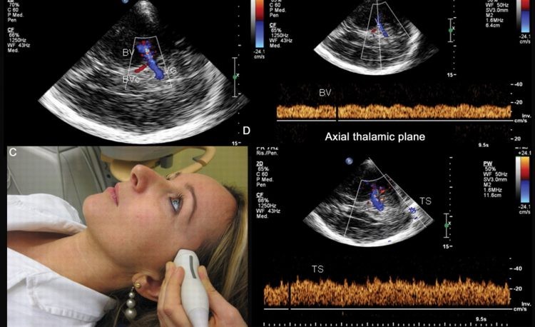
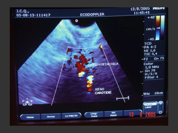
Quando e para que Fazer o Doppler Transcraniano? - Exames de Neuro

Doppler Transcraniano no Acidente Vascular Cerebral! Neuro Informa

Doppler launches the largest, national, insured, variable staff network for  Auto Dealers
Doppler launches the largest, national, insured, variable staff network for Auto Dealers

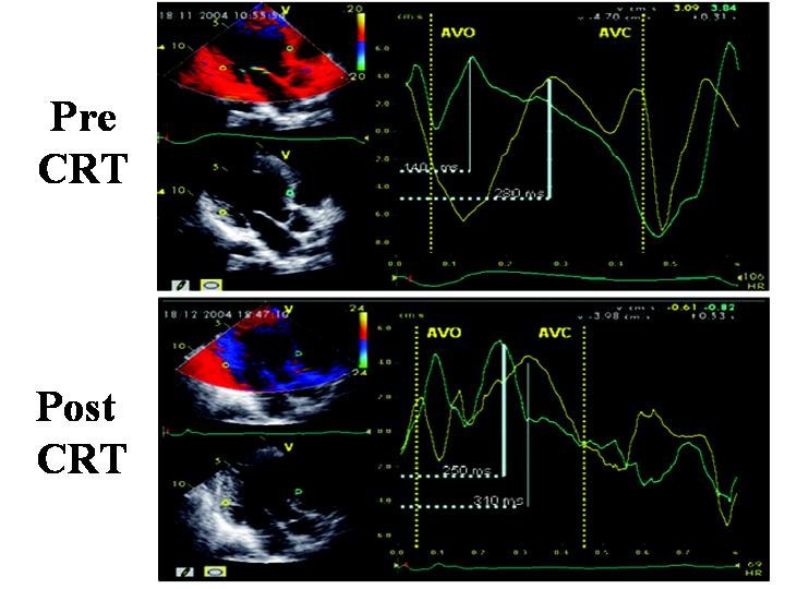
Doppler echocardiography and myocardial dyssynchrony: a practical update of  old and new ultrasound technologies | Cardiovascular Ultrasound | Full Text
Doppler echocardiography and myocardial dyssynchrony: a practical update of old and new ultrasound technologies | Cardiovascular Ultrasound | Full Text

7: Tissue Doppler-derived measurement of strain and strain Rate. During...  | Download Scientific Diagram
7: Tissue Doppler-derived measurement of strain and strain Rate. During... | Download Scientific Diagram

Serial Assessment of Left Ventricular Remodeling and Function by  Echo-Tissue Doppler Imaging After Myocardial Infarction in  Streptozotocin-Induced Diabetic Swine - ScienceDirect
Serial Assessment of Left Ventricular Remodeling and Function by Echo-Tissue Doppler Imaging After Myocardial Infarction in Streptozotocin-Induced Diabetic Swine - ScienceDirect

Ultrassom com doppler colorido das carótidas pode previnir AVC – Santa Casa  de Maceió
Ultrassom com doppler colorido das carótidas pode previnir AVC – Santa Casa de Maceió

Cardiac Time Intervals by Tissue Doppler Imaging M-Mode: Normal Values and  Association with Established Echocardiographic and Invasive Measures of  Systolic and Diastolic Function | PLOS ONE
Cardiac Time Intervals by Tissue Doppler Imaging M-Mode: Normal Values and Association with Established Echocardiographic and Invasive Measures of Systolic and Diastolic Function | PLOS ONE
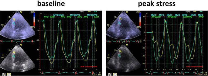
Strain Rate Imaging
Strain Rate Imaging

Tissue Doppler, Strain and Strain Rate in ischemic heart disease “How I do  it” | Cardiovascular Ultrasound | Full Text
Tissue Doppler, Strain and Strain Rate in ischemic heart disease “How I do it” | Cardiovascular Ultrasound | Full Text
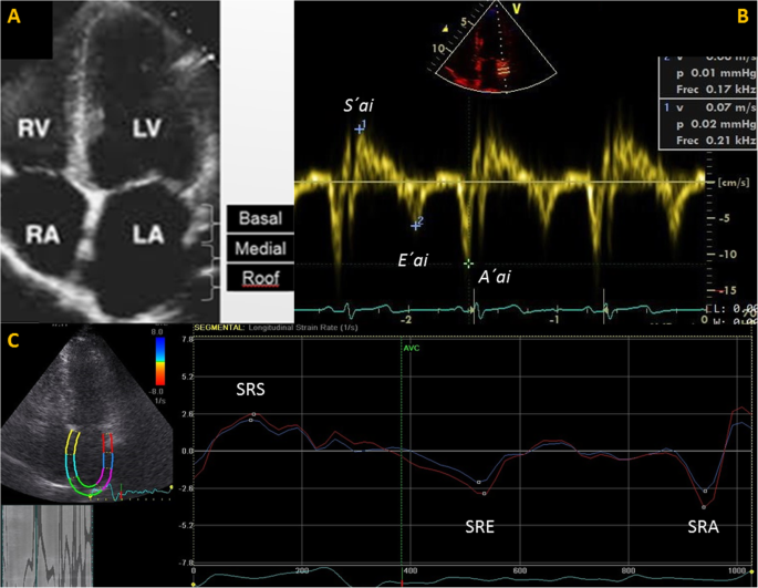
Tissue Doppler Imaging and strain rate of the left atrial lateral wall: age  related variations and comparison with parameters of diastolic function |  Cardiovascular Ultrasound | Full Text
Tissue Doppler Imaging and strain rate of the left atrial lateral wall: age related variations and comparison with parameters of diastolic function | Cardiovascular Ultrasound | Full Text

Schematic diagram of simultaneous Doppler waveforms from mitral valve... |  Download Scientific Diagram
Schematic diagram of simultaneous Doppler waveforms from mitral valve... | Download Scientific Diagram

SonoScape P20 Trolley Color Doppler Ultrasound | KeeboMed

Physiological significance of pre‐ and post‐ejection left ventricular  tissue velocities and relations to mitral and aortic valve closures -  Støylen - 2021 - Clinical Physiology and Functional Imaging - Wiley Online  Library
Physiological significance of pre‐ and post‐ejection left ventricular tissue velocities and relations to mitral and aortic valve closures - Støylen - 2021 - Clinical Physiology and Functional Imaging - Wiley Online Library

Exame reduz chances de AVC em pacientes falcêmicos

Doppler de carótidas: o exame para avaliar e prevenir o risco de AVC

Asbjørn Støylen on Twitter: "4/ and it is easily seen by transferring valve  motions from Doppler flow to Tissue Doppler recordings, where it seems that  this negative velocity is mainly septal. https://t.co/kF6Zux1DrI" /
Asbjørn Støylen on Twitter: "4/ and it is easily seen by transferring valve motions from Doppler flow to Tissue Doppler recordings, where it seems that this negative velocity is mainly septal. https://t.co/kF6Zux1DrI" /